Microscopy

About us

The Cellular Imaging Laboratory provides (fluorescence) imaging services for HUN-REN BRC researchers, for collaborators from the SZTE and other Hungarian institutes, and for EuroBioImaging members. The available equipment consists of confocal laser scanning microscopes, fluorescence and stereo microscopes. Our expertise, image processing software collection and the available courses are supporting the efficient research.

Technology

Expertise and Equipment

Leica Stellaris 8 WLL + FALCON

Our new Leica Stellaris 8 Confocal Microscope is suitable for transmission imaging (with laser scanning, non-confocal), bright field illumination and fluorescence imaging (DAPI, FITC, FITC LP, TXR cubes) with camera, confocal fluorescence imaging using white laser excitation for 5 spectral detectors. The fluorescence lifetime imaging modes (Tau sense and FALCON) are available from 440 to 790 nm excitation. Long term measurements are supported by the Adaptive Focus Control and an Okolab Incubator (temperature, CO2)

Leica SP5

The Leica TCS SP5 Confocal Microscope has 8 laser lines (405, 458, 476, 488, 496, 514, 543 and 633 nm) and 7 objectives (5x, 10x, 20x, 40x, 63x, 63x W, 25x W). AOBS (Acousto Optical Beam Splitter) is used to separate the excitation from emission light. Imaging method: scanning confocal, simulteanous 3 color detection using 3 spectral detectors plus transmission mode.

Olympus FV1000

The Olympus FV1000 Confocal Microscope has 6 laser lines (405, 458, 488, 515, 543 and 633 nm), and 6 objectives (10x/0.4, 20x/0.75, 40x/1.3 OIL, 60x/1.35 OIL, 40x/0.8 W and 60x/0.9 W). Available main dichroic beam splitters: BS20/80, DM405/488, DM/488/543/633, DM458/515 and DM405/488/543. Imaging method: scanning confocal, simultaneous 3 color detection, 2 independent spectral detectors + 1 additional PMT with emission filters + transmission mode.

VisiScope spinning disk

The Visitron VisiScope Spinning Disk Confocal Microscope has 4 laser lines (405, 488, 561 and 640 nm), and 6 objectives (4x/0.13, 10x/0.3, 20x/0.45, 40x/0.6, 60x/1.42 OIL and 100x/1.45 OIL). Detection is based on two Andor Zyla 4.2 PLUS cameras.
High speed confocal imaging, simultaneous 2 color detection and sequential multichannel acquisition available.

Olympus SZX12

The Olympus SZX12 Stereozoom Microscope has halogen transmission (bottom), LED reflection (top) and Hg fluorescence (top) illuminaton. Imaging system: Cannon 200D camera.
Available dichroic beam splitter cubes: DAPI, GFP/long pass, dsRed.

Our best

Services

3D fluorescence imaging

Confocal fluorescence multichannel high resolution image acquisition

Live cell video/time-lapse imaging

Incubation chamber with stabilized temperature and CO2 concentration

Image processing and analysis

Using ImageJ/FIJI and MATLAB, custom macros

Stereomicroscopy

Huge field of view, lower resolution; with optional fluorescence imaging

DP imaging

Differential polarization imaging, fluorescence detected anisotropy/dichroism determination, ordered molecular structures

Custom made equipment

Developing custom software and hardware solutions, IOT units

3D printing

Custom sampleholders, research tools for microscopy and more

Articles

Key publications

  • Szlanka et al. 2024
    Dominant suppressor genes of p53-induced apoptosis in Drosophila melanogaster
    G3-GENES GENOMES GENETICS Q1, https://doi.org/10.3390/ijms25074022
  • Dudits et al. 2023
    Manifestation of Triploid Heterosis in the Root System after Crossing Diploid and Autotetraploid Energy Willow Plants
    GENES, https://doi.org/10.3390/genes14101929
  • Masuda et al. 2023
    The balance between photosynthesis and respiration explains the niche differentiation between Crocosphaera and Cyanothece
    COMPUTATIONAL AND STRUCTURAL BIOTECHNOLOGY JOURNAL D1, https://doi.org/10.1016/j.csbj.2022.11.029
  • Misra et al. 2023
    Impact of protein–chromophore interaction on the retinal excited state and photocycle of Gloeobacter rhodopsin: role of conserved tryptophan residues
    CHEMICAL SCIENCE D1, https://doi.org/10.1039/D3SC02961A
  • Delawska et al. 2022
    New Insights into Tolytoxin Effect in Human Cancer Cells: Apoptosis Induction and the Relevance of Hydroxyl Substitution of Its Macrolide Cycle on Compound Potency
    CHEMBIOCHEM Q1, https://doi.org/10.1002/cbic.202100489
  • Pleckaitis et al. 2022
    Structure and principles of self-assembly of giant "sea urchin" type sulfonatophenyl porphine aggregates
    NANO RESEARCH D1, https://doi.org/10.1007/s12274-021-4048-x
  • Bernát et al. 2021
    Photomorphogenesis in the Picocyanobacterium Cyanobium gracile Includes Increased Phycobilisome Abundance Under Blue Light, Phycobilisome Decoupling Under Near Far-Red Light, and Wavelength-Specific Photoprotective Strategies
    FRONTIERS IN PLANT SCIENCE D1, https://doi.org/10.3389/fpls.2021.612302
  • Cséplő et al. 2021
    The AtCRK5 Protein Kinase Is Required to Maintain the ROS NO Balance Affecting the PIN2-Mediated Root Gravitropic Response in Arabidopsis
    INTERNATIONAL JOURNAL OF MOLECULAR SCIENCES D1, https://doi.org/10.3390/ijms22115979
  • Kaňa et al. 2021
    Fast Diffusion of the Unassembled PetC1-GFP Protein in the Cyanobacterial Thylakoid Membrane
    LIFE-BASEL Q2, https://doi.org/10.3390/life11010015
  • Radosavljević et al. 2021
    Differential Polarization Imaging of Plant Cells. Mapping the Anisotropy of Cell Walls and Chloroplasts
    INTERNATIONAL JOURNAL OF MOLECULAR SCIENCES D1, https://doi.org/10.3390/ijms22147661
  • Ünnep et al. 2020
    Thylakoid membrane reorganizations revealed by small-angle neutron scattering of Monstera deliciosa leaves associated with non-photochemical quenching
    OPEN BIOLOGY D1, https://doi.org/10.1098/rsob.200144

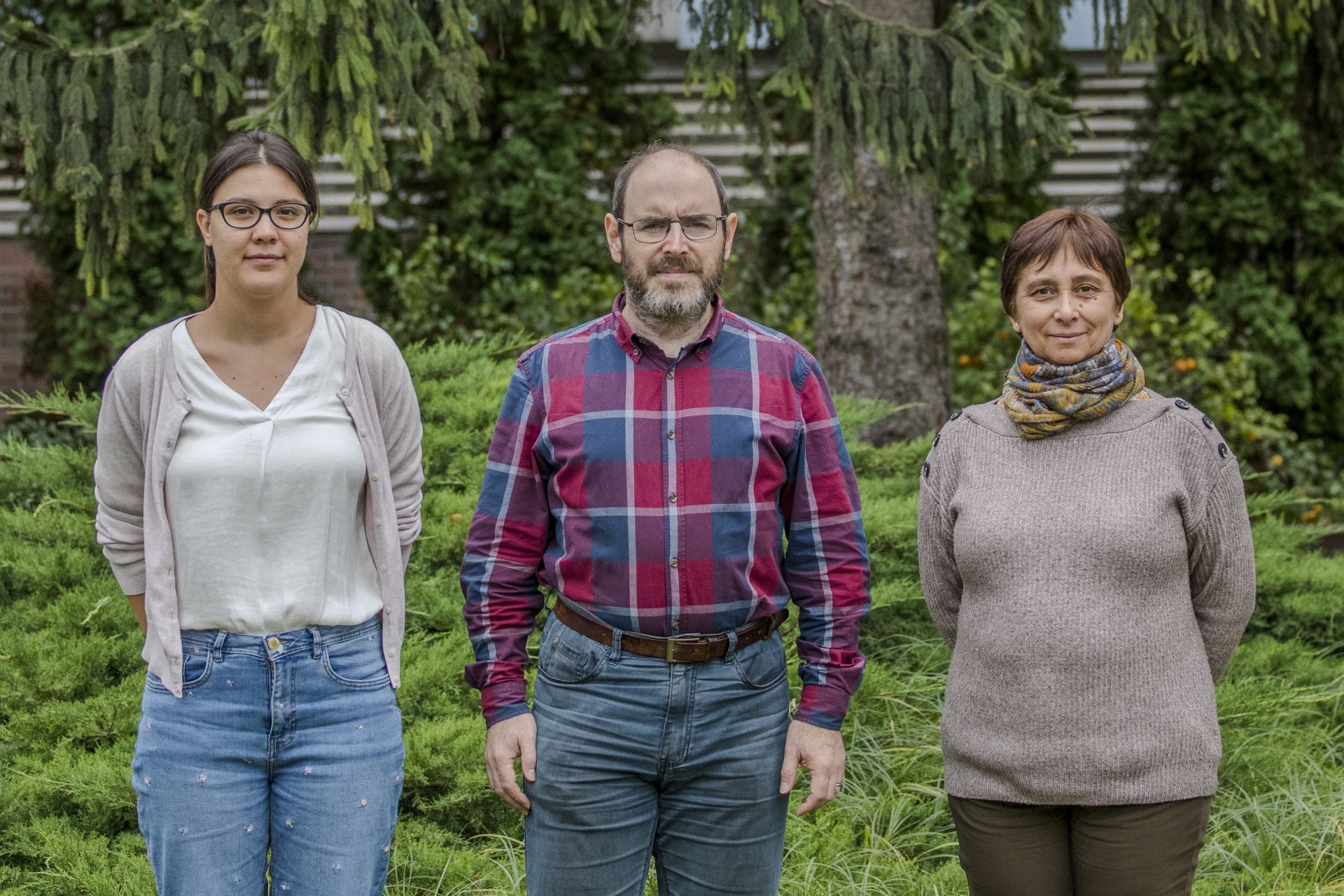
Team

Check our Team

Gábor STEINBACH

head of laboratory

Ildikó VALKONYNÉ KELEMEN

laboratory assistant/administrator expert

Lili LADÁNYI

junior research associate